SHARE

| The New York Optimist |

The Secret of Snail Patterns
Siddharth Ramakrishnan
Nature is the greatest artist and as humans we have been given the gift of creativity and hence the ability to
explore. If we take a hard look at plants and animals, we can observe how each being has been designed to all
work and fit perfectly. Artistic logic exists in the organs, cells and networks – both in their organization and
function. The amazing aspect of evolution is the common theme that progresses throughout, the commonality
across animals is very striking. The same hormone that controls reproduction in us also controls reproduction
in birds, frogs, snails and even yeast. While the variety of life forms around us is an example of adaptation and
evolution, the underlying machinery making up each organism reveals a very constant framework connecting
all life forms on a primal organic level.
At the basic level every person is an observer and so is inherently an artist as well as a scientist. The
interpretation and the medium of expression of the observations may differ but both approach the world with an
acute sense of insight and wonder. I find such wonderment in my work as a scientist, and am amazed that this
information is not abounding around, resonating with every person, each artist. I hope with this piece I can
entice you into the world of rhythmic patterns that are important to us as humans and how we can observe and
learn from such patterns that exist elsewhere in the natural world.
What do your neurons sound like? Do they burp when you burp? Do they stutter? Or is it a smooth ride like
water gushing along a sluice pipe? One can actually hear the sounds of a neuron in action by hooking it up to
a sound monitor. Electrical pulses generated by the cells can be amplified and uttered through speakers –
“Brr..up..Brr”; “Durr….Durr…Durr”, the neurons go, like a drummer performing a well practiced routine. These
rhythmic bouts are especially true of neurons that generate patterns, waves upon waves of steady activity that
convey important cyclic messages.
Such rhythmic cyclical signals are very important to us and all other animals. When you walk, you move your
left leg then your right and then the left again. This coordinated activity involves sending commands to sets of
muscles in your legs alternatively contracting and relaxing them leading to the very simple but essential action
of walking. It is a program - a program for a machine that is your body and it is centrally mediated. You don’t
have to think – “okay move left leg, then right” - you just walk. Neurons that control such rhythmic motions are
called Central Pattern Generators (CPGs) and underlie many functions like eating, locomotion, breathing in
and breathing out and many other rhythmic activities.
Studying CPGs in us is like wandering through a galaxy trying to pinpoint a clump of stars – as there are
hundreds of neurons involved. So we turn to simpler animals with fewer neurons to study such behaviors and
how they are controlled. While these animals may have fewer cells in their circuitry the principle behind their
functioning is similar to ours and we can learn a lot from observing them.
Which brings us to the protagonist of this piece – the snail. Not the garden slug with its slimy trail or the sea-
hare that grazes the ocean floor but pond snails with a maximum size of about 15mm in diameter. I had the
privilege of working with two such snails during my PhD – the Helisoma and Lymnaea (Fig. 1).

Scientist: Siddharth Ramakrishnan

Fig1. Helisoma to
the left and a pair of
Lymnaea to the right
the left and a pair of
Lymnaea to the right
Snails are wonderful creatures. It is amazing to just sit and watch them glide in the water along the glass
surface of the tank. One associates snails usually with being slow, but in water pond-snails are graceful,
cruising along effortlessly. If you let one of them wander along your finger, you may feel its tiny rasp, almost
like a miniature cats tongue licking at your skin. It is because the snails’ odontophore (or tooth-carrier, Fig2)
contains hundreds of little tiny teeth that help it to rasp at food. This brings us back to what we started with a
model system to study pattern generators. The way these snails eat is similar to us licking a scoop of ice-cream
– put out the tongue, lick at the scoop and swallow. Snails follow a similar repetitive motion while feeding
The beauty of the snail system is that the neurons that control oral movements are clumped together in the
buccal ganglion. (Note: buccal is how we refer to cheeks/oral caivity). The buccal ganglia are shaped like a
bikini, with a red background and the neurons are prominent as yellow or white polka dots of different sizes.
The arrangement of these neurons is not random – in fact in most of the snails these cells are found in a very
specific location. It is like reading a map, we can look at the buccal map and point out consistently that Neuron
A is found here, or Neuron B is located near the center etc (fig 3). We use some of the larger cells as
landmarks and orient the location of smaller cells on them. Why is this important? This is like knowing the
address of a neuron. In every snail you can look at the buccal map and say –
“Okay, that is probably Neuron A. I am going to try and record its activity.”
Recording the activity of a neuron is the next step in revealing its identity. We use a glass capillary tube with a
sharp tip (called a microelectrode) to do this. Electrical activity from the neurons is transmitted through this
electrode, through a silver wire into an amplifier. Neurons have a function – they may control muscle activity or
modulate other neurons as so they have a specific activity. Here is an example of neuron activity from snails –
The spikes are action potentials that signal excitement of the neurons and the dips indicate inhibition. Both
these signals are important and indicative of a neural pattern. And again, this activity is very consistent to a
neuron across many snails and forms a part of the neuron’s identity (like the profession of a person). The last
piece in the identity puzzle is the appearance of the neuron. We can fill a neuron with fluorescent dye and
observe how it looks (morphology). This is again consistent across most animals of one species. The location,
activity and morphology of the neuron serve as its ID – having these information makes a neuron uniquely
identifiable in every animal of that species. We have similar neurons in us, only many more making it more
difficult to establish unique identities
The other beauty of the snail system is that not only is it simple but they are also resilient. One can record from
these neurons as the snail is behaving. Based on numerous studies the CPGs underlying snail oral behavior
have been well characterized in many snail species. In both Lymnaea and Helisoma, these consist of three
phases – one controlling protraction of the tongue, one for the rasp or licking of the food and a third for
swallow. These neurons are cyclically active to produce the feeding rhythm. The duration of each part of the
cycle is modified based on how hard or soft the food is. For example if the snail is eating lettuce it will probably
take a bigger rasp than if it is on mushy watermelon – similar to how we modify our bites and chews.
.
By looking at these snail neurons we can understand how such rhythmic behaviors are modified and
modulated to precise outputs. Snails are elegant creatures whose beauty lies in their efficiency and ability to
modify their behaviors in tune with their surroundings. They are also very unique animal models where we can
look at biological phenomena from a simple cellular level to networks to whole animal behavior. They offer
insights into the machinery that makes all creatures and give us further appreciation into all the nuances that
go into such a banal but essential action such as walking or swallowing.
--
Note: This article is based on my PhD work at the University of Illinois in Chicago (2005). I worked in the
laboratory of Dr. A. Don Murphy. For a comprehensive review of snail feeding and its CPG control please refer
to – “The neuronal basis of feeding in the snail, Helisoma, with comparisons to selected gastropods.” Murphy
AD. Progress in Neurobiology. 2001, 63(4):383-408
Bio: Siddharth Ramakrishnan is a Neuroscientist currently working in the field of Bioelectronics at Columbia
University in New York. During his PhD he studied pattern generating networks in snails and how they were
modulated to elicit various behaviors. As a postdoc at UCLA (2006-09) he studied the development of neurons
in the zebrafish brain. This was accompanied by a fascination with the living world and the beauty in its
conception and patterning. He has lectured on art and science and their cusp with culture at UCLA. He is
working on a documentary with Mr. Ted Owens (Syncronous Design) on green buildings and environmental
health. Siddharth's passion lies in the integration of diverse fields and what is born at the juncture... Through
art and literature he hopes to bring in more access to science especially for children. He can be contacted at
sidramakrishnan@yahoo.com

Fig2. Scanning electron
microscope picture of snail
odontophore (or tooth
carrier). In the center each
individual square pixel is a
tooth. Scale bar is 200
micrometers.
microscope picture of snail
odontophore (or tooth
carrier). In the center each
individual square pixel is a
tooth. Scale bar is 200
micrometers.

Fig 3.The buccal Map showing
one side of the itsy-bitsy bikini.
We orient ourselves around the
ganglia using the large labeled
cells as our markers. Nerves are
indicated coming out of the
ganglia. BC indicates the
commissure that connects to a
mirror image ganglion on the
other side
one side of the itsy-bitsy bikini.
We orient ourselves around the
ganglia using the large labeled
cells as our markers. Nerves are
indicated coming out of the
ganglia. BC indicates the
commissure that connects to a
mirror image ganglion on the
other side
Fig4. Neuron activity.
Note the repetitive
pattern
Note the repetitive
pattern
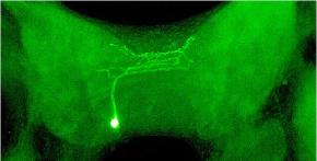
Fig 5. Morphology of a neuron filled
with Lucifer Yellow Dye
with Lucifer Yellow Dye

Fig 6. Example of rhythmic patterns.
Recording from a feeding motor neuron in
Helisoma. 1-2-3 indicates neural activity
during the three phases of feeding
Recording from a feeding motor neuron in
Helisoma. 1-2-3 indicates neural activity
during the three phases of feeding

